GEN MED BIMONTHLY ASSIGNMENT
RACHANA GANGULA
8TH SEMESTER
ROLL NO 110
I have been given the following cases to solve in an attempt to understand the topic of 'Patient clinical data analysis' to develop my competency in reading and comprehending clinical data including history, clinical findings, investigations and diagnosis and come up with a treatment plan.
Below are my answers to the Medicine Assignment based on my comprehension of the cases.
SECTION I- Pulmonology
A) “A 55 year old female patient, a resident of Miryalaguda and farmer by occupation ,chief complaints of shortness of breath, pedal edema and facial puffiness.”
Link to patient details:
https://soumyanadella128eloggm.blogspot.com/2021/05/a-55-year-old-female-with-shortness-of.html
Question 1
What is the evolution of the symptomatology in this patient in terms of an event timeline and where is the anatomical localization for the problem and what is the primary etiology of the patient's problem?
EVOLUTION OF
SYMPTOMATOLOGY

ANATOMICAL LOCATISATION-
LUNGS- COPD and Bronchiectasis
(which subsequently lead to Cardiac involvement and heart failure)
ETIOLOGY-
·
The various
reasons that could have contributed to the etiology in this patient are-
·
OCCUPATION- Patient
was a farmer by occupation. Hence, the inhalation of allergens like mold hay
spores, dust from other grains, tobacco, or some pesticides. Could also be exposure
to various other bacteria and fungi.
·
Prolonged
exposure of fumes from ‘chulha’ . Patient has been using ‘chulha’ for
the past 20 years.
Question 2
-Head elevation
- increases
hemodynamic performance and increases end expiratory lung volume
- Indications- .head injury, meningitis,pneumonia
-Antibiotics
-
INDICATIONS- severe exacerbations of COPD leading to hospitalization
-
TAB. AUGUMENTIN - broad-spectrum antibacterial
-
Formulation of Amoxicillin and Clavulanate Potassium
- TAB. AZITHROMYCIN - macrolide-type antibiotic
-Diuretics
-
INJ. LASIX IV – Furosemide
§ Loop Diuretic
§ Used to maintain blood pressure
-Corticosteroids
INJ. HYDROCORTISONE
Relieve inflammation and
decrease bronchospasm
-Nebulization
NEB. with IPRAVENT
-
Ipratropium Bromide – Bronchodilator
-
Blocking
cholinergic receptors
-
decreases
the production of cyclic guanosine monophosphate (cGMP)
-
leading
to relaxation of smooth muscles, and hence Bronchodilation
NEB. With BUDECORT
– Budesonide- corticosteroid
-
Used to control inflammation leading to bronchodilation
-Other Bronchodilators
TAB PULMOCLEAR - Acebrophylline and Acetylcysteine
- Airway mucus regulation and anti-inflammatory action causing bronchodilation
The probable causes for current acute exacerbation-
-
Triggered by an
allergen during occupational work (agriculture)
-
Triggered by environmental
pollutants
-
Underlying conditions
such as DM and HTN could have caused worsening of shortness of breath in latest
episode
-
Past history of multiple
episodes of shortness of breath which have become more frequent points to the
progression of COPD which could explain the current acute exacerbation of the
patient.
-
Improper use of
inhalational medication can also be a cause of acute exacerbation
Question 4
Could the ATT have affected her symptoms? If so how?
The various antitubercular drugs that are used in the ATT are Isoniazid,
Rifampicin, Pyrazinamide, Ethambutol, Streptomycin.
Out of these , the drugs that are known to be nephrotoxic are-
§ Rifampicin
§ Ethambutol
§ Isoniazid
They are frequently responsible for the causation of Acute Kidney
Injury.
Renal
Function Tests show increased values of both Urea as well as creatinine which
points to kidney damage.
o
Urea: 48 mg/dl
o
Creatinine: 1.9 mg/dl
Symptoms
like – Facial Puffiness and Pedal Edema could have been affected by the Acute
Injury to Kidney. Na+ and water retention is seen, leading to fluid and volume
overload, explaining the above symptoms of the patient.
Heart Failure in this patient causes decreased cardiac
output which is probably the main reason behind her electrolyte imbalance.
Hyponatremia is seen in this patient.
The association between heart failure and hyponatremia is-
NEUROLOGY-A
Link to this case - https://143vibhahegde.blogspot.com/2021/05/wernickes-encephalopathy.html
What is the evolution of the symptomatology in this patient
in terms of an event timeline and where is the anatomical localization for the
problem and what is the primary etiology of the patient's problem?
EVOLUTION OF SYMPTOMATOLOGY

ANATOMICAL LOCALISATION
ETIOLOGY
Chronic alcohol intake in this patient is the main causative factor.
Chronic alcohol intake leads to thiamine deficiency following the interference of many cellular processes which in-turn leads brain disorders like Wernicke-Korsakoff syndrome, Uremic Encephalopathy, etc.
A brief episode of alcoholic withdrawal was the cause of an episode of seizure that occurred 4 months ago. ( Alcohol withdrawal syndrome)
- Study conducted on patients with refractory partial seizures
- 96 patients received placebo (n = 96)
- 99 patients received PGB, 150 mg/day (n = 99)
- 92 patients received PGB 600 mg/day (n = 92); given 3 times a day
- Results: PGB, 150 mg/day and 600 mg/day, were both significantly more effective than placebo in reducing the RRatio ( which correspond to seizure-frequency reductions)
- Conclusions: PGB, 150 mg/day and 600 mg/day, is highly effective and well-tolerated add-on therapy in patients with partial seizures.
- randomized, double-blind trial evaluate intravenous benzodiazepines.
- Population- Adults with prolonged (lasting five minutes or more) or repetitive generalized convulsive seizures.
- Total Population- 205 patients- 66 received lorazepam, 68 received diazepam, and 71 received placebo.
- Results-status epilepticus had been terminated on arrival at the emergency department in more patients treated with lorazepam (59.1 percent) or diazepam (42.6 percent) than patients given placebo (21.1 percent)
- Conclusion- Efficacy of Lorazepam in treatement of status epilepticus is higher than both diazepam and the placebo.
- Thiamine deficiency is usually seen in chronic alcoholics mainly due to intestinal malabsorption.
- This deficiency of thiamine leads to anaerobic glucose metabolism as it is an important cofactor in various biochemical reactions.
- The lactic acid that is formed from anaerobic glycosylation leads to various pathological effects.
- It mainly affects the CNS, usually causing a presentation of characteristic symptoms of Nystagmus, Ataxia and Mental Confusion. This triad of symptoms is called Wernicke's Encephalopathywhich is commonly seen in chronic alcoholics.
- Endotoxin - induced inflammation : caused by increased gut permeability and inflammation of liver
- Myoglobin - induced tubular toxicity : due to alcoholic myopathy
- Hypoxic Tissue Injury- Decreased Renal Blood Flow : due to alcoholic cardiomyopathy.
- Hepato-Renal Syndrome in in patients with Alcoholic liver cirrhosis.
- Oxidative Stress can also lead to tissue injury of the kidneys.
- Urea: 248mg/dL (increased)
- Creatinine: 3.8 mg/dL (increased)
- Uric Acid: 18mg/dL (increased)

Question 7
Could chronic alcoholism have aggravated the foot ulcer formation? If yes, how and why?
- Yes, excessive alcohol can cause nutritional deficiencies and subsequent damage to the liver.
- These in turn can lead to decreased formation of procoagulants by the liver and alterations in the function of platelets leading to poor wound healing
- Long term excessive alcohol consumption may also lead to peripheral neuropathies which could lead to sensory nerve impairment in the foot resulting in delayed responses and cause aggravation of the ulcer.
Neurology-B
- 7 days Ago - Patient complained of giddiness. (subsided briefly; associated with one episode of vomiting)
- 4 days ago- complained of giddiness- sudden in onset, continuous and gradually progressive. It increased in severity upon getting up from the bed and while walking. Associated with bilateral healing loss and aural fullness and tinnitus.
Neurology-C

ANATOMICAL LOCALISATION
- hypokalemia (leading to palpitation)
- cervical spondylitis (causing pain the patient)
Question 2
What are the reasons for recurrence of hypokalemia in her? Important risk factors for her hypokalemia?
Question 3
What are the changes seen in ECG in case of hypokalemia and associated symptoms
Various ECG changes seen in hypokalemia are-
- decrease in T wave amplitude (earliest ECG manifestation )
- Increased P wave amplitude
- Prolongation of PR interval
- Widespread ST depression and T wave flattening/inversion
- Prominent U waves
- Supraventricular tachyarrhythmias: AF, atrial flutter, atrial tachycardia (seen on worsening of hypokalemia)
- weakness and fatigue
- muscle cramps and pain
- palpitations
- neurological symptoms- psychosis, delirium, depression
Neurology-D

QUESTION2
In the previous episodes of seizures, patient didn't lose his consciousness but in the recent episode he lost his consciousness what might be the reason?
The general occurrence of types of seizures
following Stroke in relation to their timing of occurrence after brain stroke.

*EARLY SEIZURES- [ Focal Seizures] Usually are not associated with the loss of consciousness as they are localized to one area of the cerebral hemisphere and their onset is localized.
*LATE SEIZURES- [Generalised tonic-clonic Seizures AND Complex Partial Seizures] Usually association with the loss of consciousness. These type of seizures disrupt the upper brainstem/medial diencephalon, medial and lateral fronto-parietal association cortex, which constitutes the consciousness system, hence impairing it.
Hence, the previous episodes of seizures could have been Focal Motor Seizures as they were associated with folding of fist, Tremors in leg, frothing, tongue biting, eye rolling without the impairment of consciousness
Whereas, the most recent episode being a Generalised Tonic-Clonic seizure leading to the loss of consciousness in this episode.
Another theory as to why the patient has loss of consciousness in the most recent episode of seizure is –

Neurology-E
https://nikhilasampathkumar.blogspot.com/2021/05/a-48-year-old-male-with-seizures-and.html?m=1
QUESTION 1
What could have been the reason for this
patient to develop ataxia in the past 1 year?
-
The main reason for the patient to develop
ataxia could be the effect of chronic alcohol intake on the cerebellum.
-
Direct toxicity by alcohol can lead to
cerebellar degeneration and atrophy leading to ataxia.
-
The history of chronic alcoholism and development
of ataxia point to –
WERNIKE
ENCEPHALOPATHY
A triad of symptoms of 1) Mental Confusion
2) Nystagmus 3) Cerebellar Ataxia are seen
Caused mainly in chronic alcoholics due to
thiamine deficiency
Pathology of Wernicke’s Encephalopathy

QUESTION 2
What was the reason for his IC bleed? Does Alcoholism contribute to bleeding diatheses ?
- History of Chronic alcohol intake leads to impaired platelet function and reduced platelet count which is a major cause for Intracranial Bleeding.
- Multiple head injuries due to possible minor trauma from frequent falls and no subsequent check-up can also contribute as a cause for his intracranial bleeding.
Contribution of
alcohol to bleeding diathesis

Neurology-F
QUESTION 1
Does the patient's history of road traffic accidents have any role in his present condition?
- Axonal shearing may be caused during trauma to the head
- This could aggravate the stoke
- So, yes, the history of road trauma does play a role in the condition of this patient
- Sudden confusion, trouble speaking, or difficulty understanding speech.
- Sudden numbness or weakness, especially on one side of the body.
- Sudden changes in vision
- Sudden loss of balance
- Sudden headache.
- Aspirin, antiplatelet- prevention of stroke again, to prevent other thrombotic event
- Mannitol, osmotic agent- to decrease cerebral edema, increase cerebral perfusion
- Alcohol exhibits an acute inhibitory effect on lipoprotein lipase activity which leads do decreased breakdown of lipids.
- Alcohol, hence, causes hyperlipidemia which increase in triglycerides and LDL levels.
- This inturn leads to atherosclerosis and ischemia of cerebal vessels leading to stroke

Neurology-G
- Myelopathy of hand- The changes in the hands observed in various cervical spinal disorders when there is involvement of the spinal cord.
- The main clinical features are localized wasting and weakness of the extrinsic and intrinsic hand muscles,
- Usually, there is loss of power of adduction and extension of the ulnar two or three fingers and an inability to grip and release rapidly with these fingers.
- It is a neurological sign of weak finger adduction, usually found in cervical myelopathy patients.
- It consists of involuntary abduction of the fifth (little) finger
- This is caused by unopposed action of the extensor digiti minimi.
- It is a marker for cervical compression of spinal cord.
- Procedure- Performed by tapping or flicking the nail of the middle finger and observe the response of this action on the thumb and middle finger
- Reflex flexion of thumb and index finger are signs of a positive test.
Neurology-H
What can be the cause of her condition ?
- MRI Impression in this patient- "Acute cortical vein thrombosis with hemorrhagic venous infarction involving Right posterior temporal lobe with midline shift to left by 4mm"
- Hence, cortical vein thrombosis might be the cause of her seizures.
Question 2
What are the risk factors for cortical vein thrombosis?
- Infections- Meningitis, otitis ,mastoiditis
- Prothrombotic states- Pregnancy, antithrombin deficiency, Hormone replacement therapy.
- Mechanical Injury- Head trauma, lumbar puncture
- Inflammatory- SLE, sarcoidosis, Inflammatory bowel disease.
- Nephrotic syndrome
- Drugs-Oral contraceptives, steroids, Inhibitors of angiogenesis
- Chemotherapy-Cyclosporine and l-asparginase
- Hematological-Myeloproliferative Malignancies, Primary and secondary polycythemia
- Intracranial -Dural fistula, venous anomalies
Question 3
There was seizure free period in between but again sudden episode of GTCS why?resolved spontaneously why?
Seizures are resolved and seizure free period got achieved after medical intervention but sudden episode of seizure was may be due to any persistence of excitable foci by abnormal firing of neurons.
Question 4
What drug was used in suspicion of cortical venous sinus thrombosis?
Anticoagulants are used for the prevention of harmful blood clots.
Clexane ( enoxaparin) low molecular weight heparin binds and potentiates antithrombin three a serine protease Inhibitor to form complex and irreversibly inactivates factor xa.
Cardiology-A
|
Heart
Failure with Preserved Ejection Fraction |
Heart Failure
with decreased ejection fraction |
|
Cause- Thickened
Ventricles |
Cause- Enlarged
Ventricles |
|
Also called
Diastolic Heart Failure |
Also called
Systolic Heart Failure |
|
The left ventricle
does not relax properly, resulting in impaired filling. |
This is
caused by reduced contraction of the left ventricle. |
|
Results in
increased pressure in left atria and ventricle Causing pressure
build up in the lungs. |
As a result,
not enough blood is supplied to organs. |
- Indication for doing pericardiocentesis- when pericardial effusion does not resolve on its own
- In this patient- at the time of admission pericardial fluid was 2.4mm and at the time of discharge it was 1.9 mm .
- Hence, it is showing self resolution.
- Therefore we did not do pericardiocentesis in this pt.
- age- old age
- gender- male
- hypertension
- smoking
- type 2 diabetes
- kidney disease

Cardiology-B
- Chronic alcohol abuse
- Long standing case of hypertension (19 years)
- Stage 4 Chronic Kidney disease
- ↓Erythropoietin (EPO) production by kidney due to kidney damage
- ↑ Hepcidin as a result of hepatoxicity
- Suppression of erythropoiesis as a result of circulating inflammatory cytokines released due to alcohol intake
- Hepcidin mediated decreased iron absorption from gut
- venous insufficiency
- peripheral neuropathy
- peripheral arterial occlusive disease
Cardiology-C
EVOLUTION OF SYMPTOMATOLOGY
10 YEARS AGO
- Surgery for inguinal hernia
3 YEARS AGO
- Aggravated on and off pain at the site of surgery
- On and off facial puffiness
1 YEAR AGO
- Grade II shortness of breath - diagnosed for HTN (on medication)
2 DAYS AGO
- Shortness of breath Grade II (on exertion)
- Decreased urine output
- Constipation
ONE DAY AGO
- Shortness of breath Grade IV (at rest)
DAY OF ADMISSION
- Anuria
ANATOMICAL LOCALISATION-
- Hypertension
- cholesterol levels not within healthy parameters
- Obesity
- Diabetes
- Lack of physical activity
- Age
- Smoking
Cardiology-D
(D)"67 year old patient with acute coronary syndrome"
• Heart burn like episodes since an year- relieved without medication
• Diagnosed with pulmonary TB 7 months ago- completed full course of treatment, presently sputum negative.
• Hypertension since 6 months - on medication
• Shortness of breath since half an hour-SOB even at rest
- non-surgical procedure
- uses a catheter (a thin flexible tube) to place a small structure called a stent to open up blood vessels in the heart that have been narrowed by plaque buildup ( atherosclerosis).
- Acute ST-elevation myocardial infarction (STEMI)
- Non–ST-elevation acute coronary syndrome (NSTE-ACS)
- Unstable angina.
- Stable angina.
- Anginal equivalent (eg, dyspnea, arrhythmia, or dizziness or syncope)
- High risk stress test findings.
- Intolerance for oral antiplatelets long-term.
- Absence of cardiac surgery backup.
- Hypercoagulable state.
- High-grade chronic kidney disease.
- Chronic total occlusion of SVG.
- An artery with a diameter of <1.5 mm.
- Overuse of imaging techniques such as X- RAYS AND CT SCANS as a part of routine investigations could lead to various pathological maifestations
- Overuse of imaging can lead to a diagnosis of a condition that would have otherwise remained irrelevant and could have healed naturally.
- Overdiagnosis through overtesting can also psychologically harm the patient.
- Hospitalizations or those with chronic conditions who could be treated as outpatients can lead to economic burden and a feeling of isolation.
- The use of expensive technologies and machineries can also cause a burden on health care systems.
Cardiology-E
- Atherosclerosis
- Coronary artery spasm
- Coronary artery tear
Cardiology-F
Gastro-A
EVOLUTION OF SYMPTOMATOLOGY

2) LEFT PNEUMOTHORAX SECONDARY TO BRONCHO PLEURAL FISTULA
- CBP(Complete Blood Picture ) – Usually demonstrates leukocytosis which points to an inflammatory background
- LFT(Liver Function tests )- Mainly alkaline phosphatase, total bilirubin, aspartate aminotransferase (AST), and alanine aminotransferase (ALT) levels are monitored.
- RFT(Renal Function Test )- blood urea nitrogen (BUN), creatinine, and electrolytes are monitored as their values change in light of third spacing of fluids
- Urine analysis- Urine albumins and sugars are monitored mainly to check progression of disease
- Serum amylase- significantly elevated serum amylase values are diagnostic for pancreatitis
- ABG(Arterial Blood Gas )- Mainly done when patient is in dyspnea, to identify the cause of dyspnea- ARDS or diaphragmatic irritation, etc.
- Pleural tapping – Done to assess the nature of pleural fluid as well as culture and sensitivity of the same.
- CE-CT- Various complications of pancreatitis like interstitial fluid collections, pseudocyst, necrosis etc. can be visualized.
- CXR- Done to assess extent of lung involvement.
Gastro-B
QUESTION1
What is causing the
patient's dyspnea? How is it related to pancreatitis?
- Pleural Effusion can be visualized on the Chest X-Ray report.
- This is causing the difficulty in breathing in the patient.
- The cause of Pleural Effusion can be a result of the inflammatory cytokines released in response to acute pancreatitis leading to lung damage and/or SIRS. It can also be due to fluid accumulation at base of lung due to blockage of lymphatic channels.
- Persistent dyspnea can be due to formation of sinus tract between pseudo-cyst and pleural space

QUESTION 2
Name possible reasons why the patient has developed a state of hyperglycemia?
The possible reasons to explain the state of
hyperglycemia in this patient could be-
-
Intake of large amounts of alcohol could have made the patients body more
resistant to insulin, paving the way for Diabetes Mellitus and hence explaining
the state of hyperglycemia.
-
Surges of blood glucose level are also seen in response to inflammatory
states of the body due to production of various inflammatory markers.
-
Another theory for hyperglycemic state could be the occurrence of multiple
episodes of sub-clinical acute pancreatitis leading to repeated acinar damage.
Hence causing pancreatic insufficiency and endocrine/exocrine dysfunction. This
is associated with imbalance of blood sugar regulation.
-
Initial glucagon level increase associated with defects in insulin secretion
are seen in prolonged cases of pancreatitis. This is usually associated with T3Cdm
diabetes/ brittle diabetes later in the course of the disease.
QUESTION 3
What is the reason for
his elevated LFTs? Is there a specific marker for Alcoholic Fatty Liver
disease?
·
Patient is a known alcoholic and has a
history of alcohol abuse since 4 years before he presented to the hospital.
·
While recently, his daily consumption of
locally made alcohol has signifanctly increased.
·
As alcohol is a known hepatotoxic substance,
it is associated with liver injury.
·
Hence, elevated LFT’s are a result of hepatocellular
injury due to chronic alcohol intake.
The specific markers for
Alcoholic Fatty Liver Syndrome are
-
GGT (Gamma Glutamyl Transferase)
-
CDT (Carbohydrate-deficient
transferrin)
QUESTION 4
What is the line of treatment in this patient?
1. Relieve pain in patient
-
TRAMADOL
-
ACETAMINOPHEN
2. Control Vomiting
-
ONDANSETRON
3. Control Pancreatic
Secretions
-
PANTOPRAZOLE
4. IntraVenous Fluids (
bypass GI Tract)
5. Monitor HyperGlycemia
-
Fasting and Post prandial blood glucose
-
HbA1c
6. GRBS Charting
7. USG Guided pleural tap to
control dyspnea
Gastro-C
- decreased glomerular perfusion
- decreased glomerular filtration rate
- acute renal failure.
The intermittent episodes of drowsiness could be due to Hyponatremia
Fleshy mass like casts in urine are a hallmark feature of
Acute Tubular Necrosis, hence the patient passed them in his urine.
Electrolyte Imbalances is the most probable complication
that this patient developed.
Hepatology-A
(a) "A 55 year old male patient who is a palm tree climber with PAIN ABDOMEN SINCE ONE WEEK "


Hepatology-B
- Amoebic liver abscess aspirates have an anchovy appearance
- Pyogenic abscess is usually purulent nature of aspirate . This is not the most reliable test.
- Culture of aspirate provides the most accurate diagnosis
Evolution of symptomatology with
relation to Anatomical Involvement

Evolution of symptomatology in
patient according to timeline

1)
ANATOMICAL LOCATION
Anatomical Location in this condition would primarily
involve PARANASAL SINUSES
Other anatomical parts also affected due to spread of
disease are ORBITS and CEREBRAL CORTEX
§
PARANASAL
SINUSES- predominantly, Maxillary and Sphenoid Sinus involvement is seen
– Sinusitis
§
ORBITS-
Preseptal Cellulitis
§
CEREBRAL
CORTEX- infarct around the left corona radiata
(front and temporal lobe infarcts) -right hemiparesis
2)
Primary Etiology of Patients Problem
The primary etiology surrounding this patient can be
strongly linked to a fungal infection caused by a group of opportunistic fungi
called- Mucorales.
Hence, Mucromycosis is said to be the primary cause of
disease in this patient.
the following link contains the MATERSHEET FOR COVID-19
cases-
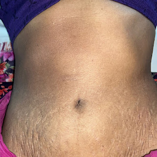

Comments
Post a Comment